43 brain mri with labels
Brain MRI: A Systematic Reading | Neurosurgery Basics An MRI will show the stroke as bright signal on the Diffusion-weighted images, and dark on the diffusion ADC sequence. An MRI is the study of choice for tumor, multiple sclerosis, and ischemic stroke. Add gadolinium contrast to evaluate tumor and abscess. For brain hemorrhage, however, CT is the go-to study. Researchers Automate Brain MRI Image Labeling Jul 28, 2021 — The researchers say it would take years to manually perform labeling of more than 100,000 MRI examinations. Deep learning typically requires ...
Brain MRI Atlas on the App Store Brain MRI Atlas is a FREE app that allows you to navigate through hundreds of of labeled brain structures. It is designed for all healthcare professionals as an interactive study and reference tool. Program Features: - Serial sequential axial T2 FLAIR images of the brain. - Structure labels organized by category.

Brain mri with labels
brain anatomy | MRI coronal brain anatomy | free MRI cross sectional ... This MRI brain coronal cross sectional anatomy tool is absolutely free to use. Use the mouse scroll wheel to move the images up and down alternatively use the tiny arrows (>>) on both side of the image to move the images.>>) on both side of the image to move the images. UCLA Brain Mapping Center - ICBM Template To view both the structural MRI and the labels launch the program typing Display icbm_template.mnc -label icbm_labels_corrected.mnc. The opacity of the labels can be set in the Colour Coding menu. The number of each label appears at the bottom left of the orthogonal views window. MRI-labeling: label human brain MRI image by AAL/BA system MRI-labeling: label human brain MRI image by AAL/BA system Under the R program environment,input an MNI coordinate, output the corresponding AAL(Automated Anatomical Labeling) and BA (Brodmann area) brain region name. More importantly, if the coordinate does not match a brain region defined by AAL/BA (e.g., white matter), the package help find the closest brain region with the corresponding ...
Brain mri with labels. Deep learning from MRI-derived labels enables automatic brain tissue ... Deep learning from MRI-derived labels enables automatic brain tissue classification on human brain CT Automatic methods for feature extraction, volumetry, and morphometric analysis in clinical neuroscience typically operate on images obtained with magnetic resonance (MR) imaging equipment. Labeled MRI Brain Scans - Neuromorphometrics We can also label scans that you provide and we are very interested in labeling white matter anatomy as seen in diffusion-weighted MRI scans. If you want an aggregate version of our data, we can provide it as a probabilistic atlas. The cost to label a single scan is $2449 (USD). Cross-sectional anatomy of the brain - e-Anatomy - IMAIOS Brain - MRI (Axial) Sagittal Coronal 3D 1/24 Revert to the old version of the viewer e-Anatomy Authors Antoine Micheau , Denis Hoa Published on Friday 15 April 2022 Section Brain DOI ISSN 2534-5079 Anatomical parts Angular gyrus Anterior cerebral artery Anterior commissure Anterior limb of internale capsule 101 labeled brain images and a consistent human cortical labeling ... given how difficult it is to label brains, the mindboggle-101 dataset is intended to serve as brain atlases for use in labeling other brains, as a normative dataset to establish morphometric variation in a healthy population for comparison against clinical populations, and contribute to the development, training, testing, and evaluation of …
Labeling Brain Structures - John Muschelli In Processing Within-Visit MRI we show how to register a T1-weighted image to the Eve template. The Eve template has two full brain segmentations and one white matter segmentations, each done by hand. I will refer to these as "atlases" because they tell you "where" you are in the brain with the corresponding labels. 1 Labels in template space Researchers automate brain MRI image labelling, more than 100,000 exams ... July 22, 2021 King's College London Researchers from the School of Biomedical Engineering & Imaging Sciences have automated brain MRI image labelling, needed to teach machine learning image recognition models, by deriving important labels from radiology reports and accurately assigning them to the corresponding MRI examinations. CaseStacks.com - MRI Brain Anatomy Labeled scrollable brain MRI covering anatomy with a level of detail appropriate for medical students. Brain: Atlas of human anatomy with MRI - e-Anatomy - IMAIOS MRI Atlas of the Brain. This page presents a comprehensive series of labeled axial, sagittal and coronal images from a normal human brain magnetic resonance imaging exam. This MRI brain cross-sectional anatomy tool serves as a reference atlas to guide radiologists and researchers in the accurate identification of the brain structures.
Researchers automate brain MRI image labeling, more than 100,000 exams ... Researchers have automated brain MRI image labeling, needed to teach machine learning image recognition models, by deriving important labels from radiology reports and accurately assigning them to... Brain lobes - annotated MRI | Radiology Case | Radiopaedia.org Head (brain-eye-ear-nose-mouth) by Razan Ali Rawashdeh. Neuro by Nicolas Moreno. Neuro- MRI by Dr Balint Botz . Smegenu_anatomija by Dr Tomas Jurevicius. Annotated CT/MR Teaching by Matt Wong . important by ESLAM DESOKY Eslam desoky abdelbadia mohammed. anatomia RMN by Joaquín Ramón Valenzuela Sánchez. Mri cnn github - domiciliotrieste.it A machine learning-based method for estimating the number and orientations of major fascicles in diffusion-weighted magnetic resonance imaging. Input image is a 3-channel brain MRI slice from pre-contrast, FLAIR, and post-contrast sequences, respectively. 8% as compared to the manually labeled ground truth. Deep learning to automate the labelling of head MRI datasets for ... Given the growing evidence that significant discrepancies can exist between labels derived from radiology reports and those derived by radiologists interrogating the actual images [21, 22], determining the validity of using report labels as proxies for image labels in the context of head MRI examinations was an important aspect of our study.
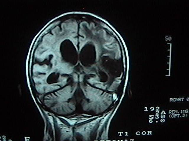
Dr Balaji Anvekar FRCR: Frontal subcortical white matter cystic lesions MRI
MRI Brain Atlas Via a toggle button, either MRI images or approximately comparable Brain Transection images may be viewed with or without labels. Navigation & Labels. The home page presents two menus for locating MRI images per transection level. In the MRI Gallery Menu, tap a reduced image to view a particular transection level.
Magnetic Resonance Imaging (MRI): Brain (for Parents) - Nemours Kidshealth
Labeled imaging anatomy cases | Radiology Reference Article ... URL of Article. This article lists a series of labeled imaging anatomy cases by body region and modality. On this page: Article: Brain. Head and neck. Spine. Chest. Abdomen and pelvis.

BIOLOGY BLOG, : July 2012
MRI Brain Animated Quiz - University of Minnesota Note: spacebar toggles labels; also arrow keys do Previous/Next Sequentially click/tap: first the dot associated with a term; then, its corresponding target dot on the MRI image. If a line connection appears, your choice was correct!
Some sample MRI images
Brain MRI: How to read MRI brain scan | Kenhub MRI is the most sensitive imaging method when it comes to examining the structure of the brain and spinal cord. It works by exciting the tissue hydrogen protons, which in turn emit electromagnetic signals back to the MRI machine. The MRI machine detects their intensity and translates it into a gray-scale MRI image.

Brain Differences in College Aged Occasional Drug Users - Neuroscience News
NITRC: Manually Labeled MRI Brain Scan Database: Tool/Resource Info Manually Labeled MRI Brain Scan Database. Visit Website . Image 1 of 3 Click for more. This is a continuously growing and improving database of high-quality neuroanatomically labeled MRI brain scans, created not by an algorithm, but by neuroanatomical experts. All results are checked and corrected.

Brain MRI - Neurology Photo (6815446) - Fanpop
High resolution automated labeling of the hippocampus and amygdala ... We trained a 3D deep CNN to label the hippocampus and amygdala on whole brain 700 μm isotropic 3D MP2RAGE MRI acquired at 7T. Manual labels of the hippocampus and amygdala were used to (i) train the predictive model and (ii) evaluate performance of the model when applied to new scans.
Magnetic Resonance Imaging (MRI) – RadLink
101 Labeled Brain Images and a Consistent Human Cortical ... by A Klein · 2012 · Cited by 650 — An automated labeling system for subdividing the human cerebral cortex on MRI scans into gyral based regions of interest. Neuroimage 3, 968–980.
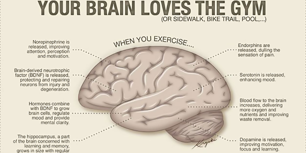
Exercise Can Help Increase Size Of The Brain’s Hippocampus
Labels · Aytijha/Brain-MRI-Alzheimers-Prediction · GitHub A Deep Learning approach to use Brain MRI scans to detect Alzheimer's Disease - Labels · Aytijha/Brain-MRI-Alzheimers-Prediction
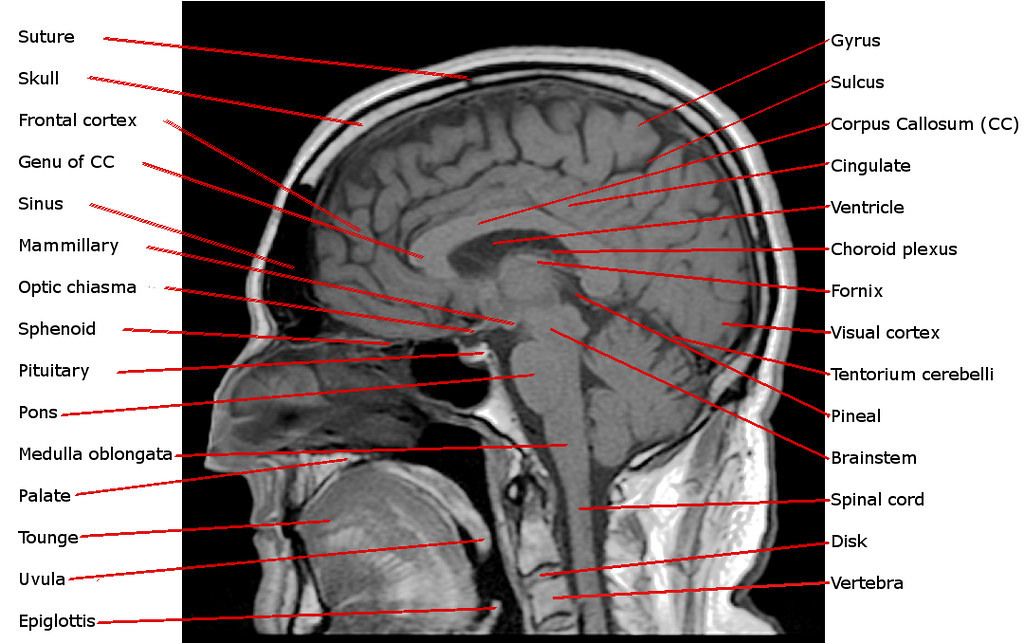
Annotated Sagittal T1 Midline MRI Scan of Reigh's Brain | Flickr
Automatic anatomical brain MRI segmentation combining label propagation ... Both the simulated data and experimental values of SI were fitted to the model of Eq. . We explored the variation of the parameters a and b as functions of the standard deviations used for the simulated data (σ sys and σ rand).Using the brain label data, we determined the variation of a and b depending on the registration method employed (unregistered, rigid propagated, affine propagated ...

MRI Procedure of Brain
Automatic Structural Parcellation of Mouse Brain MRI Using Multi-Atlas ... Compared to human brain MRI segmentation studies , , , the availability of mouse brain atlas databases is lacking, and as such, the performance of label fusion techniques is subsequently limited. To the best of our knowledge, there are currently only two in vivo multi-atlas mouse brain MRI databases that are publicly available [3] , [5] .

Brain Radiology | 127.164
How to Read an MRI: 15 Steps (with Pictures) - wikiHow An MRI machine uses a magnetic field to produce detailed images of the brain, spine, heart, bones, and other tissue. Most modern MRI centers can give you a copy of your MRI on a disc or flash drive after your appointment. While only your...

MRI Brain Planning
Atlas of BRAIN MRI - W-Radiology Brain magnetic resonance imaging (MRI) is a common medical imaging method that allows clinicians to examine the brain's anatomy (1). It uses a magnetic field and radio waves to produce detailed images of the brain and the brainstem to detect various conditions (2).

MRI BLOG: Brachial Plexus MRI (II/II)
MRI anatomy | free MRI axial brain anatomy MRI anatomy | free MRI axial brain anatomy This MRI brain cross sectional anatomy tool is absolutely free to use. Use the mouse scroll wheel to move the images up and down alternatively use the tiny arrows (>>) on both side of the image to move the images.

MzTeachuh: Special Needs Tweets of the Day 6/24/14
MRI-labeling: label human brain MRI image by AAL/BA system MRI-labeling: label human brain MRI image by AAL/BA system Under the R program environment,input an MNI coordinate, output the corresponding AAL(Automated Anatomical Labeling) and BA (Brodmann area) brain region name. More importantly, if the coordinate does not match a brain region defined by AAL/BA (e.g., white matter), the package help find the closest brain region with the corresponding ...

Anatomy Course - Labelled Quizzes | Radiopaedia.org
UCLA Brain Mapping Center - ICBM Template To view both the structural MRI and the labels launch the program typing Display icbm_template.mnc -label icbm_labels_corrected.mnc. The opacity of the labels can be set in the Colour Coding menu. The number of each label appears at the bottom left of the orthogonal views window.

Brain and Spines: Diffusion and hypoglycemia
brain anatomy | MRI coronal brain anatomy | free MRI cross sectional ... This MRI brain coronal cross sectional anatomy tool is absolutely free to use. Use the mouse scroll wheel to move the images up and down alternatively use the tiny arrows (>>) on both side of the image to move the images.>>) on both side of the image to move the images.

Brain MRI Cross Section 4
Post a Comment for "43 brain mri with labels"