40 diagram of the lungs with labels
en.wikipedia.org › wiki › AcetylcholinesteraseAcetylcholinesterase - Wikipedia Acetylcholinesterase (HGNC symbol ACHE; EC 3.1.1.7), also known as AChE, AChase or acetylhydrolase, is the primary cholinesterase in the body. It is an enzyme that catalyzes the breakdown of acetylcholine and some other choline esters that function as neurotransmitters. Lungs (Human Anatomy): Picture, Function, Definition ... The lungs are a pair of spongy, air-filled organs located on either side of the chest (thorax). The trachea (windpipe) conducts inhaled air into the lungs through its tubular branches, called...
Label the lung diagram Diagram - Quizlet Start studying Label the lung diagram. Learn vocabulary, terms, and more with flashcards, games, and other study tools.

Diagram of the lungs with labels
Can you label the lungs? Quiz - PurposeGames.com This is an online quiz called Can you label the lungs? There is a printable worksheet available for download here so you can take the quiz with pen and paper. From the quiz author. Labeling the lungs. This quiz has tags. Click on the tags below to find other quizzes on the same subject. lungs. respiratory system. Lung diagram Images, Stock Photos & Vectors | Shutterstock 8,489 lung diagram stock photos, vectors, and illustrations are available royalty-free. See lung diagram stock video clips Image type Orientation Sort by Popular Healthcare and Medical Anatomy Recreation/Fitness Diseases, Viruses, and Disorders lung respiratory system medicine pulmonary alveolus human body organ Next of 85 Label Lungs Diagram Printout - EnchantedLearning.com ... This is part 6 of a 7 part hands-on unit study on anatomy of the human body. Create a lung model, measure lung capacity, dissect a lung, play a respiratory relay race, and more! These lessons are geared toward 4th-5th grade level children and their siblings. They were created by another creative mom for our weekly homeschool co-op.
Diagram of the lungs with labels. Diagram Of The Respiratory System With Labels Stock Photos ... Browse 154 diagram of the respiratory system with labels stock photos and images available, or start a new search to explore more stock photos and images. Newest results The respiratory system Lungs with Alveoli Labeled The digestive system lung. Human Respiratory System Lungs Label Design Anatomy Human Lungs Bronchitis - Wikipedia Bronchitis is inflammation of the bronchi (large and medium-sized airways) in the lungs that causes coughing.Symptoms include coughing up sputum, wheezing, shortness of breath, and chest pain.Bronchitis can be acute or chronic.. Acute bronchitis usually has a cough that lasts around three weeks, and is also known as a chest cold. In more than 90% of cases the cause … Labeled diagram of the lungs/respiratory system. - SERC View Original Image at Full Size. Labeled diagram of the lungs/respiratory system. Image 37789 is a 1125 by 1408 pixel PNG Uploaded: Jan10 14. Last Modified: 2014-01-10 12:15:34 Label the Lungs Diagram | Quizlet ... superior lobe of right lung ... middle lobe of right lung ... inferior lobe of right lung ... superior lobe of left lung ... left main (primary) bronchus ... lobar (secondary) bronchus ... segmental (tertiary) bronchus ... inferior lobe of left lung ... Sets found in the same folder Bi 233: Labeling the Larynx 21 terms SunshineGirl79 the cell
› labelling_interactives › 1Label the heart - Science Learning Hub Jun 16, 2017 · Labels. Description. Vena cava. Carries deoxygenated blood from the body to the heart. Semilunar valve. Flaps that prevent backflow of blood. Left atrium. Receives oxygenated blood from the lungs. Left ventricle. Region of the heart that pumps oxygenated blood to the body. Pulmonary artery. Carries deoxygenated blood to the lungs. Right ventricle › photos › human-throat-anatomyHuman Throat Anatomy Stock Photos, Pictures & Royalty-Free ... Human Respiratory System anatomical vector illustration, medical education cross section diagram with nasal cavity, throat, lungs and alveoli. Human Respiratory System anatomical vector illustration, medical education cross section diagram with nasal cavity, throat, esophagus, trachea, lungs and alveoli. human throat anatomy stock illustrations Label Lungs Diagram Printout - EnchantedLearning.com Read the definitions below, then label the lung anatomy diagram. bronchial tree - the system of airways within the lungs, which bring air from the trachea to the lung's tiny air sacs (alveoli). cardiac notch - the indentation in the left lung that provides room for the heart. diaphragm - a muscular membrane under the lungs. Fully Labelled Diagram Alveolus Lungs Showing Stock Vector ... Find Fully Labelled Diagram Alveolus Lungs Showing stock images in HD and millions of other royalty-free stock photos, illustrations and vectors in the Shutterstock collection. Thousands of new, high-quality pictures added every day.
Lung Anatomy, Function, and Diagrams - Healthline The lungs begin at the bottom of your trachea (windpipe). The trachea is a tube that carries the air in and out of your lungs. Each lung has a tube called a bronchus that connects to the trachea.... Lobes of the Lung - SmartDraw Lobes of the Lung Lobes of the Lung Create healthcare diagrams like this example called Lobes of the Lung in minutes with SmartDraw. SmartDraw includes 1000s of professional healthcare and anatomy chart templates that you can modify and make your own. 4/22 EXAMPLES EDIT THIS EXAMPLE Text in this Example: Lobes of the Lung Anatomy of the Lung | SEER Training Anatomy of the Lung. The lungs are the major organs of the respiratory system, and are divided into sections, or lobes.The right lung has three lobes and is slightly larger than the left lung, which has two lobes.. The lungs are separated by the mediastinum.This area contains the heart, trachea, esophagus, and many lymph nodes. The lungs are covered by a protective membrane known as the pleura ... Labeled Diagram of the Human Lungs - Bodytomy Given below is a labeled diagram of the human lungs followed by a brief account of the different parts of the lungs and their functions. Each lung is enclosed inside a sac called pleura, which is a double-membrane structure formed by a smooth membrane called serous membrane.
The Bronchia | ClipArt ETC
› consumers › consumer-updatesConsumer Updates | FDA Science-based health and safety information you can trust.

Cartilaginous Fish: Unique Characteristics
qualifications.pearson.com › content › damInternational GCSE Biology - Edexcel May 23, 2013 · 1 The diagram shows part of a food web in an oak forest. (a) Use the information in the food web to complete the statements in the table. The first one has been done for you. (4) Statement Number the number of animals is 8 the number of producers is the number of herbivores is the number of secondary consumers is the number of food chains is deer

Circulatory System Diagram - Cardiovascular System and Blood Circulation Diagram
diagram of how blood flows through the heart - Lisbdnet.com 28.11.2021 · Blood comes into the right atrium from the body, moves into the right ventricle and is pushed into the pulmonary arteries in the lungs. After picking up oxygen, the blood travels back to the heart through the pulmonary veins into the left atrium, to the left ventricle and out to the body’s tissues through the aorta.. How does blood flow through the heart step by step quizlet?

Respiratory System Information and Labeled Diagrams Flashcards | Quizlet
byjus.com › biology › human-heartHuman Heart - Anatomy, Functions and Facts about Heart The right ventricle pumps the blood to the lungs for re-oxygenation through the pulmonary arteries. The right semilunar valves close and prevent the blood from flowing back into the heart. Then, the oxygenated blood is received by the left atrium from the lungs via the pulmonary veins. Read on to explore more about the structure of the heart.

How to draw Biology diagrams
Lung Diagram Labelling Activity | Primary Resources | Twinkl This handy Lung Labelling Worksheet gives your children the opportunity to show how much they've learned about the human lung system. The beautifully hand-drawn illustration shows a lung diagram, labelled with blank spaces where learners can fill in its different components.
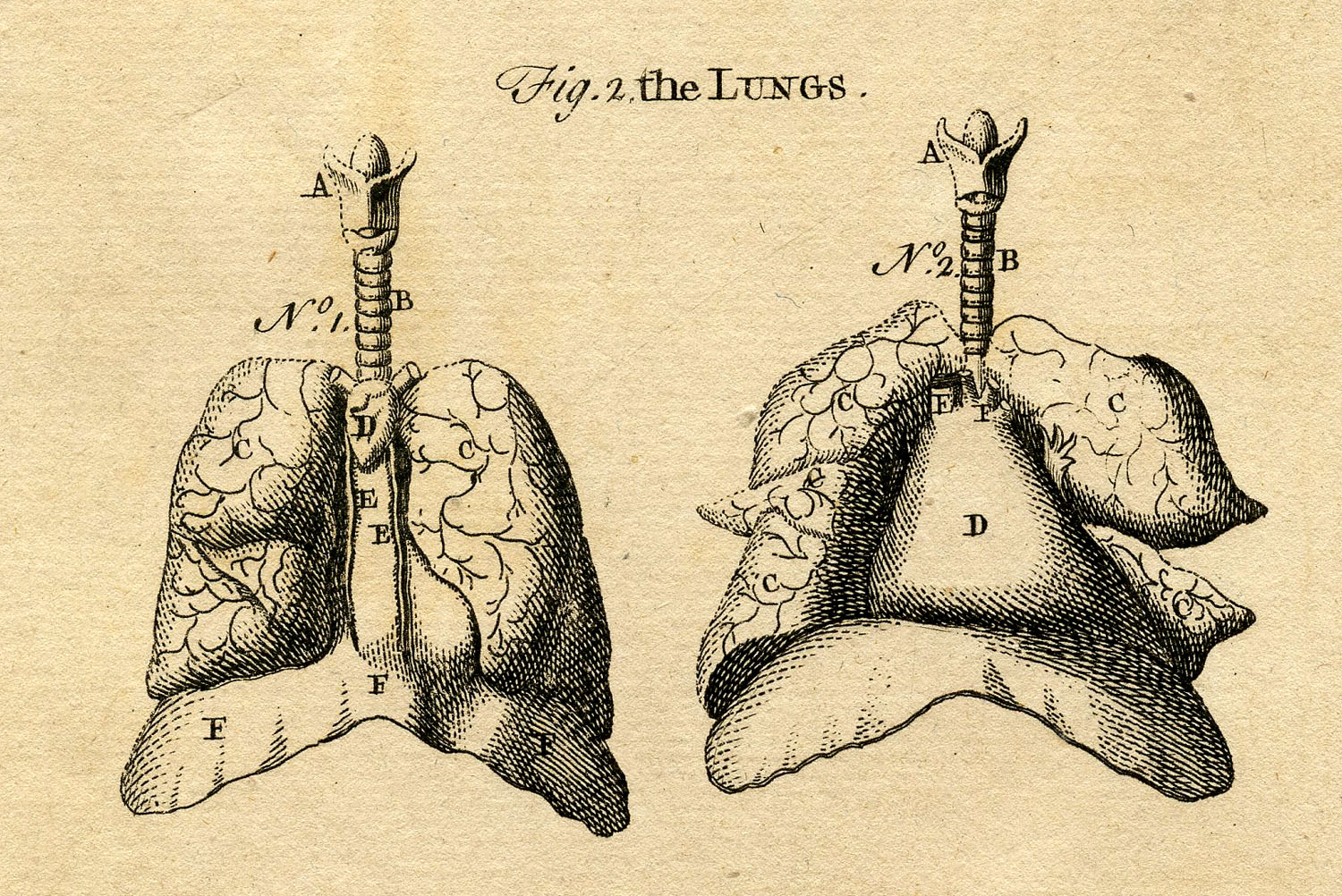
Early Anatomy Graphics - Diagram of Lungs - The Graphics Fairy
Normal CT chest lung on axial images with labels | e ... Normal anatomy of the thorax on labeled chest CT with scrollable images: radiological anatomy in axial slice of the lungs, mediastinal lymph nodes, trachea, bronchi, pleural cavity, heart and pulmonary vessels.

your lungs and how breathing works diagram of the human respiratory system infographic by ross ...
A Guide to Understand Lung with Diagrams | EdrawMax Online To learn the lung anatomy and its function, the students can use the lung diagram. As it can be tough to create a lung diagram by hand, the students can use the EdrawMax Online tool. 2. The Lungs Anatomy How does the lung look like, or what is the structure of lung. In this part, it will give you a detailed introduction of lung structure.
Post a Comment for "40 diagram of the lungs with labels"